

A page from the "Causes of Color" exhibit...
How do different colorblind individuals see?

Many people mistakenly think anyone labeled as "colorblind" only sees black and white - like a black and white movie or television show. On the contrary, it is extremely rare to have monochromacy - the complete absence of any color sensation. The variations in type and severity of colorblindness are explored in more detail in the section Colorblindness.
|
|
   
|
|
Comparison of normal and dichromat vision |
||
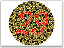
Anomalous trichromacy (where vision is based on three functioning cone types, but one or more cone types are atypical) is by far the most common form of colorblindness, and in many senses it is the least severe. Its effect is to reduce the ability to discriminate between colors, but it does not eliminate color perception altogether. People with this form of colorblindness experience very little difficulty in doing tasks that require color vision. Some may not even be aware that their color perception is in any way atypical. The only problem they have is passing color vision tests.
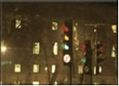
Under poor viewing conditions, such as when driving in dazzling sunlight or in rainy or foggy weather, it is easily possible for protanomalous individuals to mistake a blinking red traffic light for a blinking yellow or amber one, or to fail to distinguish a green traffic light from the various "white" lights in store fronts, signs, and street lights. The dimming can be so pronounced that reds may be confused with black or dark gray, and red traffic lights may appear to be extinguished.
Dichromats (those with vision based on two functioning cone types) on the other hand, can be so severely color deficient as to affect their daily lives. The real problem, for protanopes (absence of L-cone), deuteranopes (absence of M-cone) and tritanopes (absence of S-cone), is there are far too many color names and no obvious basis for using one instead of another. Why call something "orange" when it doesn’t look different in any way from something else called green, tan, beige, or any of several other color names?
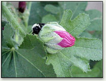
For the protanope, red, orange, yellow, yellow-green, and green, appear somewhat shifted toward green in hue, and all appear paler than they do to the average observer. Protanopes may learn to distinguish reds from yellows and from greens primarily on the basis of their apparent brightness or lightness, rather than any perceptible hue difference. Violet, lavender, and purple are indistinguishable from various shades of blue because their reddish components are so dimmed as to be invisible. Pink flowers, reflecting both red light and blue light, may appear blue to the protanope. In the pictures below, the orange flowers completely disappear.
The deutanope suffers the same hue discrimination problems as the protanope, but without the dimming. The names red, orange, yellow, and green mean very little. Similarly, violet, lavender, purple, and blue appear to be the same color. In the pictures below, the orange flowers completely disappear.
Tritanopes can have difficulty distinguishing green, cyan, and blue. Tritanopes can also have problems distinguishing yellow from violet. They can have pink, orange, and brown confusions. In the picture below, the orange and pink flowers look the same.
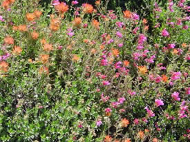
Typical |
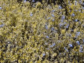
Protanope (L-cone absent) |
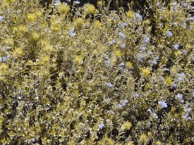
Deutanope (M-cone absent) |
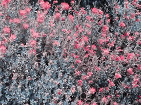
Tritanope (S-cone absent) |
|
Comparison of normal and dichromat vision |
|
The vision simulator illustrates these differences more clearly.
Achromatopsia and monochromacy
Achromatopsia (sometimes called rod monochromacy) can be inherited (congenital), or cerebral achromatopsia can develop later in life. Because an achromat or achromatope’s vision comes entirely from the rods, it works best at low light levels, and at the periphery of view. People with the condition must wear dark sunglasses in daylight or bright indoor conditions. Congenital achromats perceive black, white, and shades of grey, but have no understanding of the concept of color. People with cerebral achromatopsia see in shades of grey, because having had color vision, they perceive the absence of color.
There are three types of cone monochromacy (all of which are rare), named according to the working cone type, e.g. blue cone monochromacy is a condition in which the L-cones and M-cones are missing, leaving just the S-cones and the rods. Because the S-cones do not contribute to our perception of brightness, blue cone monochromats have similar problems with bright lights to achromatopes, although they are able to distinguish a small range of colors. Monochromats can see during the day, but can’t distinguish between hues.